Analysis of the application value of 3D printing technology in ankle surgery
Summary
3D printing technology has the advantages of rapid, individualized, intuitive and operability in the field of ankle surgery. The ankle orthosis made with 3D printing technology can achieve individualized treatment and shorten the production time; pre-synthesis of bone 3D printing model and guiding model can reduce the difficulty of surgery and intraoperative perspective, and enhance the accuracy of surgery; 3D before surgery Print technology to produce individualized bone fragments and prostheses for better matching. This article reviews the application value of 3D printing technology in ankle surgery.
The foot and ankle has the characteristics of many bones and joints, complex shape and small volume. Therefore, the diagnosis and treatment of trauma in this part has always been one of the difficulties in the field of orthopedics. With the rapid development of computer technology, 3D printing technology came into being. Thanks to the rapid and individualized advantages of 3D printing technology, some research difficulties in the foot and ankle area have been broken. This article reviews the application status of 3D printing technology in the field of ankle surgery.

1.3D printing technology overview
The generalized 3D printing technology, also known as rapid prototyping technology , is a technology based on a digital model that constructs objects by layer-by-layer printing under computer control. Objects of any geometric shape can be constructed using this technique. Since Hull et al. made the first 3D printer in 1986, this technology has been developed for 30 years. After entering the 21st century, due to the rapid upgrade of computer technology, the efficiency and performance of 3D printers have made great progress. The output and sales volume have risen rapidly, the price has been decreasing year by year, and even the basic home 3D printer has appeared. Nowadays, more and more powerful new 3D printing technologies are emerging. These new 3D printers can print more items with different materials and different needs. The development of 3D printing technology has made the application of 3D printers more and more extensive, and various industries have begun to explore the application value of 3D printers in this field.
2.3D printing steps and methods
3D printing is mainly divided into 3 steps. The first step is to make a 3D design on the computer, that is, to create a 3D model, then partition or slice the 3D model so that it can be recognized by the 3D printer; the second step is done by the printer, that is, to read the partition or slice of the 3D model. The data is printed and then the partitions or slices are bonded into a whole by various means; the third step reprocesses the second printed out of the body to make it more suitable for the actual needs, thus completing the entire printing process. In the field of clinical medicine, modern imaging techniques such as multi-slice spiral CT and MRI can provide morphological parameters for modeling 3D printing. With the gradual regularization of these tests in the clinic, 3D printing has begun to have application space in the medical field.
At present, the 3D printing technology applied in medicine mainly includes selective laser sintering, fused deposition modeling, multi-nozzle molding technology and stereo printing. The selective laser sintering technique refers to selective cutting of a thermoplastic sheet by a laser beam, thereby forming each layer of CT or MRI into a thermoplastic sheet; and then laminating the sheets to form a 3D model. The model produced by this technology has the advantage of high geometric precision, but its disadvantage; however, the high cost and rough surface make it less practical. The fused deposition modeling technique refers to layering and solidifying a molten material. This method allows different tissues to be made of different materials, and each anatomical structure can be made of materials of different colors and types to distinguish them. The geometric accuracy and surface quality of the model are high, but it takes a long time, and the model can take up to 24 hours. As the earliest 3D printing technology, stereo printing is still more commonly used in the medical field. The principle is that the liquid photosensitive resin to be formed is selectively polymerized under the action of a laser, and hardened and laminated to form a model. The finished model has the advantages of sterilizable, high geometric precision, high surface quality and high level of detail. Its detail level and surface quality exceed the fused deposition modeling technology, but it also has the disadvantage of long time. Multi-nozzle molding technology is called narrow-range 3D printing technology. The printing method is similar to that of a common inkjet printer. The powdered material is sprayed layer by layer to form a finished model. This printing method has many disadvantages, but it takes less time and has lower material costs. If the latest technology is used to make the finished product, the time can be compressed to within 4 hours, and there is still room for shortening.
Application of 3.3D printing technology in ankle surgery
3.1 personalized foot and orthosis customization
The ankle orthosis is a device used to control the movement of the ankle and foot and can be used to fix arthritis, fracture sites and correct ankle and ankle deformities. Mass-produced orthose models are limited and do not offer individualized services for optimal orthopedic effects. Although the customized ankle orthosis solves the above problems, its production takes a long time and requires high technical skills. The 3D printed ankle orthosis uses reverse engineering and rapid prototyping technology to achieve the same or even better results than the customized ankle orthosis, while greatly reducing the difficulty of making the individualized ankle orthosis and shortening the production time. The ankle orthosis made by Faustini and others using laser sintering technology has a personalized shape and function, and performs well in terms of resistance to pressure and torsion. Mavroidis et al. collected individualized data of patients through laser scanning, and used 3D printing technology to make models. The finished products were similar to the traditional polypropylene orthoses in terms of stretchability, elasticity, bending resistance and compression resistance. Creylman and other applications of laser sintering technology have customized new ankle orthoses for 8 patients who have used custom ankle orthoses for more than 2 years. As a result, walking experiments have shown that these ankle orthoses perform well, at stride distances, Both the stride time and the start time have achieved the same good results as the custom ankle orthosis, and the time to make these ankle orthoses (only 1d) is much shorter than the custom ankle orthosis. At present, the ankle orthosis used in 3D printing has performed well in various tests and experiments, and it is expected to be put into clinical application.
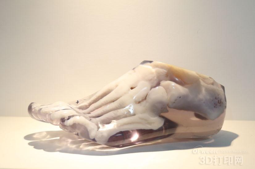
3.2 Preparation of the foot skeleton model
The foot and ankle have many bones and complex shapes. The surgeon needs to have solid anatomical knowledge, strong spatial imagination, rich clinical experience and perfect preoperative design. Even with the above conditions, patient individualized differences can lead to increased difficulty in surgery and increased operating time. Preparing a 3D printed model of the ankle bone before surgery can provide a realistic simulated surgical environment to help the surgeon to fully observe and understand the surgical site. In addition, the 3D print model of the ankle bone can also be used for pre-pre-reset, intraoperative plate screw placement and internal fixation simulation, and can pre-form the steel plate to further adjust the surgical plan to determine a more reasonable and individualized The internal fixation is placed in position and angle for better surgical results and shorter intraoperative time.
Kacl et al. compared the 3D printed model of the foot and ankle bone of 30 cases of intra-articular fractures with common 2D and 3D reconstruction images. It was found that there was no significant difference between the 3D printed model and the common 2D and 3D reconstructed images in the diagnosis of calcaneal fractures. Compared with the digital virtual model, the 3D printing model has greater operability, so it is more practical in surgical design. Giovinco et al. introduced the use of 3D printing technology to assist in the treatment of Charcot foot, which is to use a common 3D printer to make several inexpensive bone models of the foot. The surgeon can effectively reduce the difficulty of Charcot foot surgery and improve the operation efficiency, thus reducing the operation efficiency. The risk of surgery increases the success rate of surgery. Bagaria et al. used a 3D printing technique to create a 16-year-old male patient with a calcaneal fracture (Sanders IIB) model. The model clearly showed the fracture; the surgery was successfully designed, and the patient recovered well after 2 years. Completely healed. Chung et al reported a 3D printed model for unilateral intra-articular calcaneal fracture surgery, which first scanned the CT image of the bilateral calcaneus, and then applied the mirror technique to print the contralateral calcane as the same as the affected calcaneus. The size of the model, using the model and imaging data to design the position of the steel plate, the nail path, etc., so that the internal fixation can be stably fixed, and can be placed with a small incision, and the steel plate is pre-shaped, so that It can better fit the bone surface; the model is compared with the fracture under direct vision and X-ray fluoroscopy. Based on this, the reduction, bone grafting and fixation are performed, and the final operation is successful. Zhang Ying et al. studied the effect of computerized rapid prototyping technology on individualized treatment of trigeminal fractures, and compared with conventional surgery, it was found that although there was no difference in the exposure time between the two, the former had a shorter reduction and fixation time, and the operation was excellent. The rate is also higher. Jin Dan et al. have prepared a navigation template for the lower jaw combined separation and fixation by reverse engineering method and three-dimensional reconstruction technology. The prepared guide template has good matching, and it is used in the clinical application of the lower jaw combined with internal fixation. Shows good results. Bone 3D printing model and guiding template can help fracture reduction, plate pre-bending, internal fixation placement and angle determination, osteotomy orthopedic design, etc., because it can reduce the difficulty of surgery, increase the accuracy of surgery, and reduce intraoperative X-ray The number of fluoroscopy uses has a more optimistic application prospect in clinical practice.
3.3 Personalized design of the implant
Injury to the ankle and foot often causes bone defects, and proper bone grafting is required to reshape the structural integrity. However, due to the complex bone structure of the ankle and the skeletal defect, the shape of the implant often takes a lot of time during the operation. Using the 3D printing model of the ankle bone, the individualized bone fragments and prosthesis can be prepared by applying 3D printing technology before surgery, which not only saves the operation time but also achieves better matching than the traditional method. Due to technical limitations, the direct printing of titanium alloy steel plates or screws using 3D printers is still in the experimental stage. At present, 3D printing technology is mainly used for pretreatment of steel plates. The steel plates treated by 3D printing technology can be more individualized to adapt to the patient's bone condition than traditional steel plates. Thereby achieving better results. Cooke et al. applied degradable biomaterials in 3D printing to successfully produce bone fragments that meet the requirements. Leukers et al. used 3D printing technology to print hydroxyapatite scaffolds and culture stem cells thereon to make these scaffolds resemble human bone structures. 3D printing technology can construct objects of any shape, so its application value in ankle surgery is worthy of further study.
4. Summary and outlook
The ankle orthosis made with 3D printing technology can not only achieve individualized treatment, but also reduce labor and time costs. For complex ankle surgery, 3D printed models and guided templates not only reduce the difficulty of surgery, reduce the number of intraoperative perspectives, but also enhance the accuracy of surgery. In addition, 3D printing technology can also be applied to the manufacture of individual implants. With advances in 3D printing model surface treatment and geometric accuracy, 3D printing technology has a huge space for direct printing of implants. However, there are still some shortcomings in current 3D printing technology. First of all, although the cost of 3D printing model has been steadily decreasing year by year with the advancement of technology, it is still relatively high. Secondly, 3D printing is still too long and difficult to apply to emergency surgery. Finally, the application of 3D printing technology is still in use. In the experimental phase, the scope of application needs more exploration, and the actual efficacy needs to be verified by clinical research. If 3D printing technology can be continuously improved and continue to develop, its application prospect in ankle surgery will be very broad.
References (omitted)
cat toys interactive,cat toys wand,cat toys balls,cat teething toys
AUTRENDS INTERNATIONAL LIMITED , https://www.petspetscare.com