Swiss team pioneers low-power laser 3D printing technology with potential for surgical applications
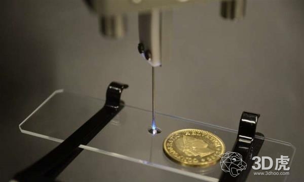
Recently, Swiss researchers have made new breakthroughs in the field of biological 3D printing . They developed an endoscopic SLA technique that uses ultra-fine fibers to focus the laser beam to create a very small-scale structure. This innovative approach can one day be used to print biocompatible structures directly into tissue in the body, with the potential to repair damage and a range of other critical applications.

Currently, there are a number of laser-based 3D microfabrication technologies on the market, but most of the methods rely on complex laser equipment, which may be too expensive and too bulky. These methods utilize an optical phenomenon known as two-photon polymerization, while the new method utilizes different phenomena in which solidification of a particular chemical occurs only above a certain threshold of light intensity.   

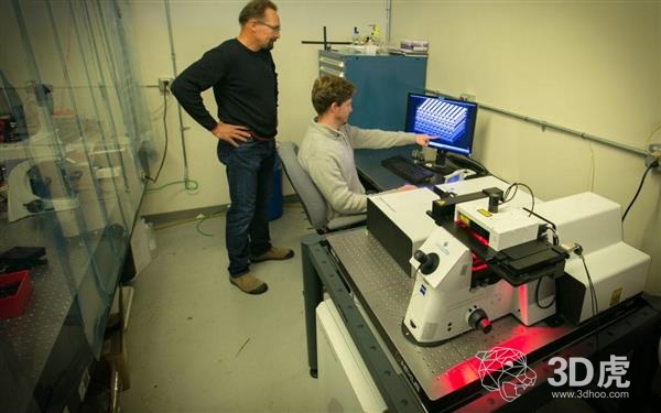
(two-photon laser equipment)
According to Paul Delrot, head of the research group at the Federal Institute of Technology in Lausanne, Switzerland, “We have the expertise to operate and shape light through fiber optics, which allows us to think that microstructures can be printed in a compact system. In addition, in order to make the system more affordable, we use non- Linear dose-responsive photopolymers, which can be operated with simple continuous wave lasers, so no expensive pulsed lasers are needed.

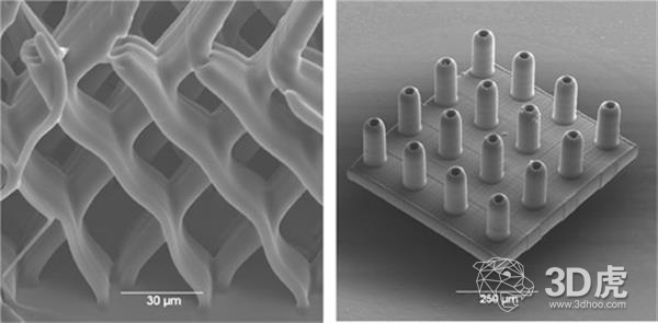
(Structure made using two-photon polymerization)
The relatively inexpensive continuous laser beam used by the team emits light at a wavelength of 488 nm. This is in the visible range and is safer for human cells than other types of lasers. They focus the beam through a tiny fiber that specifically cures specific areas of the droplets of the photosensitive liquid. This is similar to the stereolithography 3D printing method, but at a much smaller scale. The photopolymer used is a combination of an organic polymer precursor and a photoinitiator made of a chemical which is moderately priced and readily available.
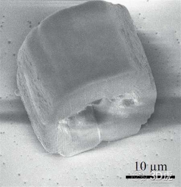
The laser device is calibrated prior to the microfabrication process to focus the light without having to move the fiber. Hollow and solid microstructures are highly accurately formed in the photopolymer, with a 1 micron lateral (one side to the other) print resolution and a 21.5 micron axial (depth) print resolution. The success of this approach means that it can be quickly used to study how cells interact with various microstructures in animal models, ultimately paving the way for human endoscopic 3D printing.

(structure created using new technology)
“With further development, our technology can make endoscopic micromanufacturing tools valuable in the surgical process. These tools can be used to print microscopic or nanoscale three-dimensional structures that promote cell adhesion and growth to create engineering. Organize and restore damaged tissue,†Delrot said. “Compared to the most advanced two-photon photopolymerization system, our equipment has a rougher print resolution, but studying cell interactions is potential because our approach is not Requires complex optical components and can therefore be applied to current endoscope systems."
It is reported that the research results have been published in the "OpticsExpress" journal. The next phase of the research team was to develop a new biocompatible photopolymer chemical during the surgical implementation of their pioneering technology. They also need to create a compact delivery system for this material. Another requirement is faster scanning speeds, but this limitation can be overcome by using commercial endoscopes rather than ultra-thin fibers because the size of the instrument is not necessarily relevant for all applications. The team also needed to develop a technology to complete and post-process the in vivo printed microstructure to make it suitable for biomedical functions.
Classified Bin,Recycling Pedal Trash Can,Modern Soft Close,Recycle Trash Can
Qin Jian International Limited , https://www.luanbosmart.com